

Cardiac Stress Tests: Types, Methods, and What Results Mean for Heart Health
Heart disease remains the leading cause of death worldwide, making early detection and accurate diagnosis critical. Among the tools physicians use to evaluate heart health, cardiac stress testing is one of the most valuable. These tests allow providers to see how the heart performs under physical or chemical “stress,” revealing abnormalities that may not appear when the body is at rest.
In this comprehensive guide, we’ll cover:
-
What cardiac stress tests are and why they’re ordered
-
The different ways “stress” can be induced (treadmill exercise vs. medications)
-
The different ways heart performance can be assessed (EKG, echocardiography, nuclear imaging, and others)
-
Specialized stress test options
-
What results mean and how they guide treatment
What Is a Cardiac Stress Test?
A cardiac stress test is a diagnostic tool that evaluates how well the heart responds to increased demand. The test is designed to increase the workload on the heart—either through physical activity (such as walking on a treadmill) or with medications that simulate exercise.
By doing this, stress tests can reveal:
-
Blockages in coronary arteries that limit blood flow
-
Abnormal heart rhythms triggered by exertion
-
Poor pumping function of the heart muscle
-
Exercise tolerance and fitness capacity
Stress testing is especially useful because many people with heart disease may have normal test results while at rest, but symptoms and abnormalities only appear during activity or increased workload.
Why Do Doctors Order Stress Tests?
Cardiac stress tests are not one-size-fits-all. Physicians order them for several reasons, including:
1. Diagnosing Coronary Artery Disease (CAD)
CAD occurs when fatty plaques build up inside coronary arteries, restricting blood flow. Stress testing can reveal whether reduced blood supply causes chest pain (angina) or EKG changes during exertion.
2. Evaluating Chest Pain or Shortness of Breath
When patients present with unexplained symptoms, stress tests help determine if the cause is heart-related.
3. Assessing Heart Rhythm Disorders
Exercise or medications can trigger arrhythmias that are not present at rest.
4. Determining Exercise Capacity
For patients undergoing rehabilitation or athletes returning to play, stress tests provide objective measurements of functional capacity.
5. Guiding Treatment Decisions
Stress test results can inform whether a patient needs medication adjustments, stenting, bypass surgery, or simply lifestyle modification.
6. Monitoring Heart Health After Procedures
Stress testing can help assess how well a stent, bypass surgery, or other cardiac intervention is performing.
How Stress Is Induced: Exercise vs. Medication
To challenge the heart, a stress test must raise heart rate and increase demand for oxygen. This can be accomplished in two main ways:
1. Exercise Stress (Treadmill or Bicycle Test)
-
Patients walk on a treadmill or pedal a stationary bike while the intensity gradually increases.
-
Speed and incline are raised every few minutes (commonly using the Bruce protocol).
-
The test continues until the patient reaches target heart rate, develops symptoms, or shows concerning findings.
Advantages:
-
Mimics real-life exertion
-
Provides data on exercise capacity
-
Allows evaluation of blood pressure and heart rate responses
Limitations:
-
Not possible for patients with mobility issues, orthopedic limitations, or severe illness
2. Pharmacologic (Medication-Induced) Stress
For those unable to exercise adequately, medications are used to simulate stress. Common options include:
-
Adenosine or Regadenoson: Dilate blood vessels, mimicking exercise by increasing blood flow differences between healthy and blocked arteries.
-
Dobutamine: Increases heart rate and contractility, similar to exercise.
Advantages:
-
Allows testing for those who cannot exercise
-
Can be combined with imaging for precise results
Limitations:
-
May cause side effects such as flushing, headache, palpitations, or shortness of breath
How Heart Performance Is Assessed During Stress Testing
Once stress is induced—either through exercise or medication—the next step is to assess how the heart responds. Several modalities are used, each with unique benefits.
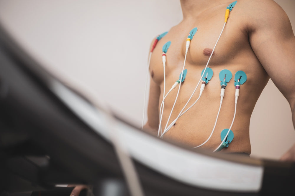
1. Exercise Stress Test with EKG (Electrocardiogram)
This is the simplest and most widely used form of stress testing.
-
Electrodes are placed on the chest to monitor the heart’s electrical activity during exercise.
-
Physicians look for changes in ST-segments, arrhythmias, or abnormal blood pressure responses.
What it detects:
-
Evidence of coronary artery disease (ischemia)
-
Exercise-induced arrhythmias
-
Fitness and functional capacity
Advantages:
-
Inexpensive and widely available
-
Non-invasive
-
Provides quick results
Limitations:
-
Less sensitive than imaging-based tests
-
May miss mild blockages
-
Requires adequate ability to exercise
2. Stress Echocardiography
This test combines stress induction (exercise or dobutamine) with ultrasound imaging of the heart.
-
Images are taken before and immediately after stress.
-
Doctors compare wall motion to see if parts of the heart fail to contract normally under stress (a sign of reduced blood flow).
What it detects:
-
Ischemia due to blocked arteries
-
Heart muscle viability
-
Valve function under stress
Advantages:
-
More sensitive than EKG alone
-
No radiation exposure
-
Provides detailed structural information
Limitations:
-
Operator-dependent (image quality may vary)
-
Difficult in patients with obesity or lung disease
3. Nuclear Stress Test (Myocardial Perfusion Imaging)
This advanced test uses small amounts of radioactive tracers (such as technetium or thallium) and a special gamma camera to create images.
-
Images are taken at rest and after stress.
-
Areas with reduced blood flow show up as “cold spots.”
What it detects:
-
Areas of reduced or absent blood flow
-
Severity and extent of coronary artery disease
-
Heart muscle viability
Advantages:
-
Highly accurate and sensitive
-
Provides information on prognosis
-
Useful in patients with prior heart surgery or stents
Limitations:
-
Radiation exposure
-
More expensive than EKG or echo
-
Requires specialized equipment
4. Cardiac MRI Stress Test
Cardiac MRI with pharmacologic stress (often using adenosine or dobutamine) provides highly detailed images of both blood flow and heart muscle.
Advantages:
-
No radiation
-
Superior soft-tissue imaging
-
Can assess both structure and function
Limitations:
-
Limited availability
-
Expensive
-
Not suitable for patients with certain implants or severe claustrophobia
5. PET Stress Test (Positron Emission Tomography)
PET stress testing is similar to nuclear imaging but uses more advanced tracers, offering higher accuracy and lower radiation exposure.
Advantages:
-
Very high sensitivity and specificity
-
Better for obese patients or those with lung disease
Limitations:
-
Very limited availability
-
Higher cost
6. Stress Testing with Cardiopulmonary Exercise Testing (CPET)
This test combines treadmill or bicycle exercise with measurement of oxygen consumption (VO2 max) and carbon dioxide output.
Advantages:
-
Best test for measuring exercise capacity and endurance
-
Useful in evaluating unexplained shortness of breath
-
Helps distinguish between cardiac and pulmonary causes of symptoms
Limitations:
-
Requires specialized equipment and expertise
Interpreting Stress Test Results
Stress test results are interpreted in the context of the patient’s symptoms, history, and risk factors.
Normal Results:
-
No evidence of ischemia
-
Adequate blood flow and heart function under stress
-
Normal heart rate and blood pressure responses
Abnormal Results:
-
ST-segment changes on EKG
-
Wall motion abnormalities on stress echo
-
Reduced tracer uptake in nuclear or PET scans
-
Reduced ejection fraction or pumping capacity under stress
Risks and Safety of Stress Testing
While stress tests are generally safe, they do carry small risks:
-
Arrhythmias: Exercise or medications may trigger abnormal heart rhythms
-
Chest Pain or Heart Attack: Rare, but possible in patients with severe disease
-
Side Effects of Medications: Flushing, headache, nausea, or shortness of breath
Stress testing is always conducted under close medical supervision with emergency equipment available.
Choosing the Right Stress Test
The best type of stress test depends on:
-
The patient’s ability to exercise
-
Clinical suspicion of coronary artery disease
-
Previous heart surgeries or interventions
-
Need for detailed imaging
-
Patient safety and radiation exposure concerns
Typical approach:
-
Start with an exercise EKG stress test if the patient can exercise and has no baseline EKG abnormalities.
-
Use stress echo or nuclear stress test if more detail is needed.
-
Consider PET or MRI stress tests for complex cases or when other tests are inconclusive.
Conclusion
Cardiac stress testing remains one of the most powerful tools in cardiology, helping physicians uncover hidden coronary artery disease, assess heart function, and guide treatment. Whether performed with a treadmill, bicycle, or medication, and assessed with EKG, echo, nuclear imaging, MRI, or PET, stress testing provides essential information about heart health.
If you have symptoms such as chest pain, shortness of breath, or unexplained fatigue, or if you’re at high risk for heart disease, talk to your healthcare provider about whether a stress test may be right for you.
Call to Action
At Revolution Health, we believe in proactive heart health care. If you’re concerned about your heart or want to know which cardiac test is right for you, schedule an evaluation with our team. Together, we’ll design a personalized plan to protect your heart and optimize your long-term health.
References
-
Gibbons RJ, Balady GJ, Bricker JT, et al. ACC/AHA 2002 Guideline Update for Exercise Testing. Circulation. 2002.
-
Armstrong WF, Zoghbi WA. Stress Echocardiography: Current Methodology and Clinical Applications. J Am Coll Cardiol. 2005.
-
Henzlova MJ, Duvall WL, Einstein AJ, et al. Stress protocols and tracers. J Nucl Cardiol. 2016.
-
Greenwood JP, Maredia N, Younger JF, et al. Cardiovascular magnetic resonance and single-photon emission computed tomography for diagnosis of coronary heart disease (CE-MARC). Lancet. 2012.
-
Taqueti VR, Di Carli MF. Coronary Microvascular Disease Pathogenic Mechanisms and Therapeutic Options. J Am Coll Cardiol. 2018.